Client: Professor Michael Corrin
Year: 2019
Media: Horos, Cinema 4D, Adobe Illustrator, Adobe Photoshop
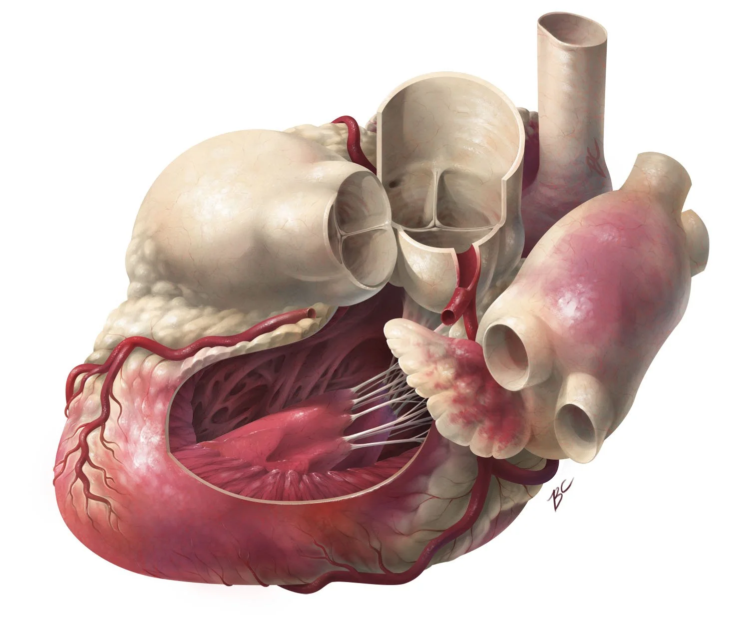
Anatomical illustrations usually depict standard views and cross-sections. The goal of this piece was to depict an organ from an unusual angle and with an unusual cut, which required 3D assets and a great deal of research. This posterolateral view of the heart shows the interior of the left ventricle, including the papillary muscles, tendinous cords, and trabeculae carnae, as well as the pulmonary and aortic cusps.
Process Work
Deciding the View and Cut
Using Horos, I extracted a rough 3D model of the heart. This model included the coronary arteries (not emphasized), left auricle (grey), pulmonary cusps (also grey), aortic cusps (red), mitral valve (green), and papillary muscles (blue and dark teal). Then, I imported the model into Cinema 4D to determine which view and cut would show the structures of interest most effectively.
Preliminary Studies
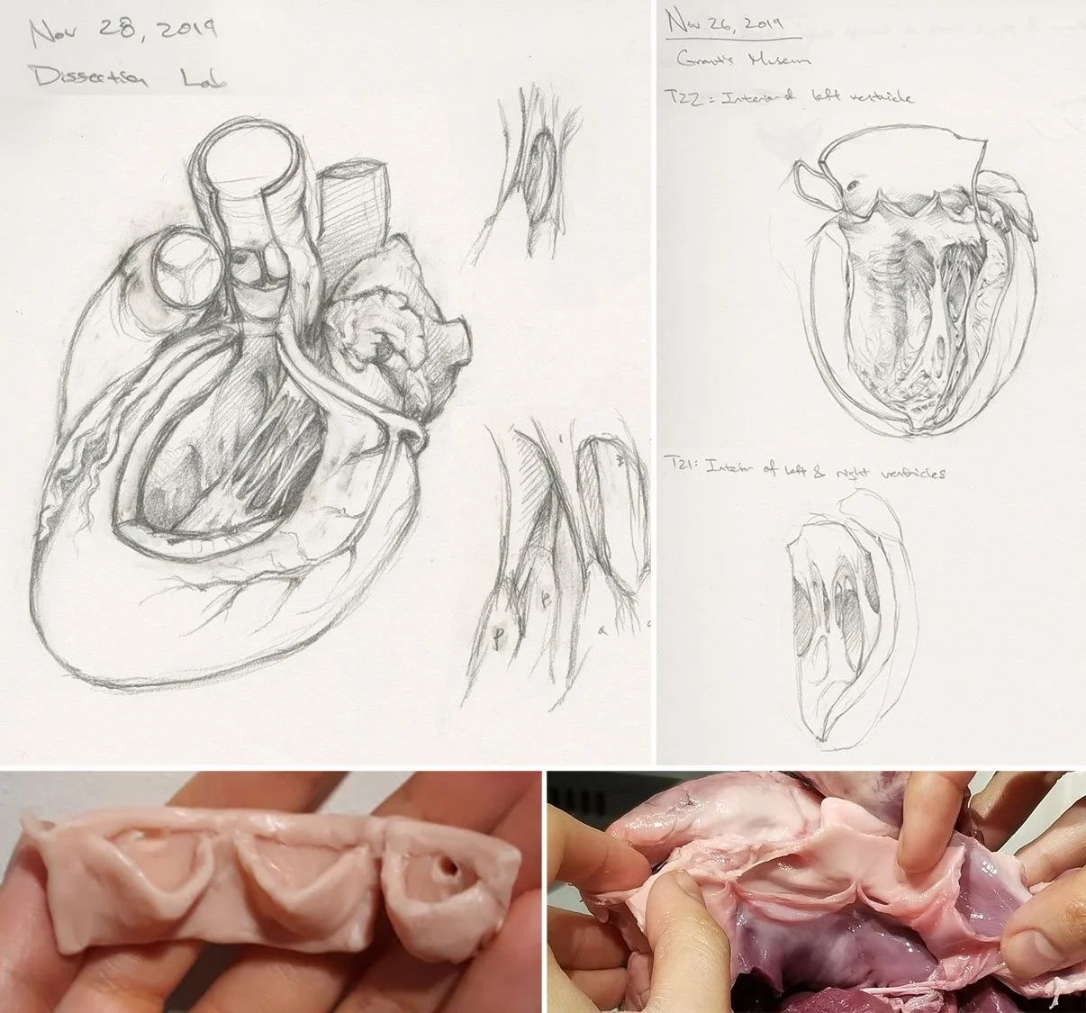
To understand the finer details that the rough 3D model was unable to capture, I sketched a dissected cadaver’s heart and specimens from Grant’s museum, modeled the aortic cusps using clay, looked at a pig’s heart, and referenced illustrative and photographic atlases. This helped with things such as wall thickness, the trabeculae carnae, the cusps, and the mitral valve.
Sketch Process
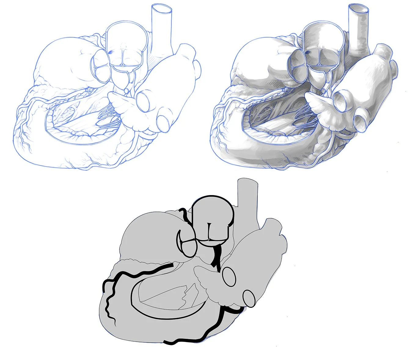
Using the 3D model of the heart as a base and ensuring details were accurate using my other references, I created the sketch and added preliminary shading in Adobe Photoshop. Then, I blocked out the shapes in Adobe Illustrator.
Rendering Process
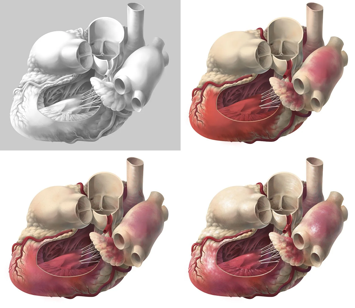
Using Adobe Photoshop, I finished rendering the heart in greyscale. I added colour and texture using layers set to multiply mode, subtle colour variation using colour mode, and specular highlights with pure white on normal mode.
References
Academic Sources
Agur, Anne M. R., and Arthur F. Dalley. 2019. Moore’s Essential Clinical Anatomy. 6th ed. Philadelphia: Wolters Kluwer.
Anderson, Robert H., Diane E. Spicer, Anhony M. Hlavacek, Andrew C. Cook, and Carl L. Backer. 2013. Wilcox’s Surgical Anatomy of the Heart. 4th ed. Cambridge: Cambridge University Press.
Desai, Milind, Christine Jellis, and Teeerapat Yingchoncharoen. 2015. An Atlas of Mitral Valve Imaging. London: Springer.
Dal-Vianco, Jason P., and Robert A. Levine. 2013. “Anatomy of the mitral valve apparatus: role of 2D ad 3D echocardiography.” Cardiology Clinics 31 (2): 151-164. Doi: 10.1016/j.ccl.2013.03.001
Ho, Siew Yen. 2009. “Structure and anatomy of the aortic root.” European Journal of Echocardiography 10 (1): i3-i10. doi: 10.1093/ejechocard/jen243
Johannes Sobotta. 2001. Sobotta Atlas of Human Anatomy Volume 2: Trunk, Viscera, Lower Limb, edited by Reinhard Puz and Reinhard Pabst. 13th ed. Philadelphia: Loppincott Williams & Wilkins.
Pernkopf, Eduard. 1943. Topographische Anatomie des Menschen. Volume 1, 2nd half. Berlin: Urban & Schwarzenberg.
Rohen, Johannes W., and Chihiro Yokochi. 1993. Color Atlas of Anatomy: A Photographic Study of the Human Body. 3rd ed. New York: Igaku-Shoin.
Saremi, Farhood, Stephan Achenbach, Eloisa Arbustini, and Jagat Narula. 2011. Revisiting Cardiac Anatomy: A Computed-Tomography-based Atlas and Reference. 1st ed. Oxford: Wiley-Blackwell
Visual References
Al Jazeera America. “Transporting a beating heart for transplant – TechKnow”. YouTube. Accessed December 9, 2019. https://www.youtube.com/watch?v=vZNa_I4xBnk
Cadaver’s heart from MSC1001 Dissection Laboratory. University of Toronto. Accessed November 28, 2019.
Grant’s Museum. “T21: Interior of left and right ventricles” and “T22: Interior of left ventricle”. University of Toronto. Accessed November 26, 2019.
Emilie Roudier. “Promising Research Shows Blood Vessel Growth Key to Healthy Fat Tissue.” York University. Accessed December 10, 2019. https://news.yorku.ca/files/Screen-Shot-2018-12-04-at-10.15.47-AM-1024x582.png
Joni Subroto. “AMAZING Living Human Heart in Box! Medical Breakthrough 2017.” YouTube. Accessed December 9, 2019. https://www.youtube.com/watch?v=XyEHJUUs-RA
Pig’s heart from First Choice Supermarket. 7866 Kennedy Road, Markham. Accessed December 13, 2019.
Pixmeo. “AGECANONIX”. OsiriX: DICOM Image Library. Accessed November 18, 2019. https://www.osirix-viewer.com/resources/dicom-image-library/


