Client: Professor David Mazierski
Year: 2020
Media: Adobe Illustrator, Adobe Photoshop
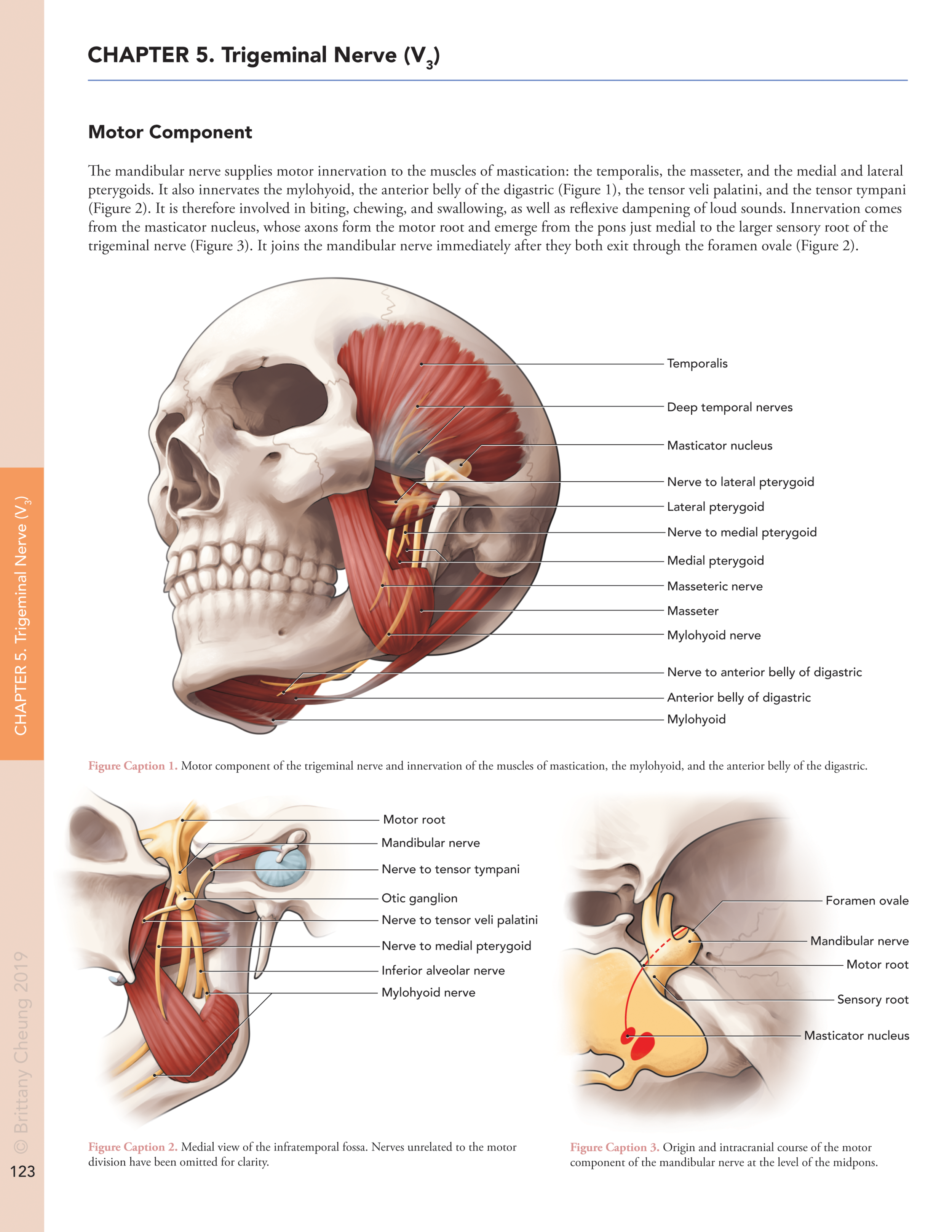
The trigeminal nerve is the largest and most complex cranial nerve. This textbook page is intended to teach neuroanatomy students about its motor component, which is found in its third division, the mandibular nerve.
References
Academic Sources
Binder, Devin K. 2010. Cranial Nerves: Anatomy, Pathology, and Imaging (Binder, Devin K., Christian Sonne, D., and Fischbein, Nancy J, Eds.). New York: Thieme Medical Publishers. Pages 47-51. Figures 5.1: Intracranial course of the trigeminal nerve, and 5.5: Mandibular nerve (CN V3).
Spalteholz, Werner. 1903. Hand Atlas of Human Anatomy Vol. III: Viscera, Nerves, Sense-Organs. Philadelphia: J. B. Lippincott. Pages 686-697, Fig. 761: Passage of nerves through the dura mater and the skull, Fig. 762: Right ganglion semilunare [Gasseri], Fig. 763: Nerves of the right orbital cavity, viewed from above, 1st layer, Fig 765: Nerves of the right orbital cavity and of the upper jaw, and Fig. 771: Right ganglion oticum, viewed from within.
Standring, Susan (Ed.). 2008. Gray’s Anatomy: The Anatomical Bases of Clinical Practice, 40th ed. London: Churchill Livingstone. Pages 276, 287, 433, and 542. Fig. 19.2: Ventral aspect of the brain stem, Fig. 19.16: The trigeminal nerve and its central connections, Fig. 27.11: The innervation of the cranial meninges, and Fig. 31.15: Arteries and nerves of the head, deepest lateral region.
Williams, Peter L. and Warwick, Roger (Eds.). 1980. Gray’s Anatomy, 36th ed. London: Churchill Livingstone. Page 1065, Fig. 7.181: The right otic ganglion and its branches displayed from the medial side.
Wilson-Pauwels, Linda, Akesson, Elizabeth J., and Stewart, Patricia A. 1988. Cranial Nerves: Anatomy and Clinical Comments. Hamilton, Ontario: B.C. Decker Inc. Pages 49-59, Figures V-1: Overview of Trigeminal Nerve, V-2: Ophthalmic Division of Trigeminal Nerve, V-3: Trigeminal Nucleus (Dorsal View of Brain Stem), V-5: Branchial Motor Component of Trigeminal Nerve, and V-6: Medial Aspect of the Lateral Wall of the Mandible.
Visual References
Jario, Martín. 2015. Skull downloadable. Sketchfab. Accessed on February 22, 2020. https://sketchfab.com/3d-models/skull-downloadable-1a9db900738d44298b0bc59f68123393
what-when-how. n.d. Brainstem II: Pons and Cerebellum Part 2. The-Crankshaft Publishing. Accessed on February 29, 2020. http://what-when-how.com/neuroscience/brainstem-ii-pons-and-cerebellum-part-2/ Fig. 11-5: Middle pons.

