Client: Professor David Mazierski
Year: 2019
Media: Adobe Photoshop, Adobe Illustrator
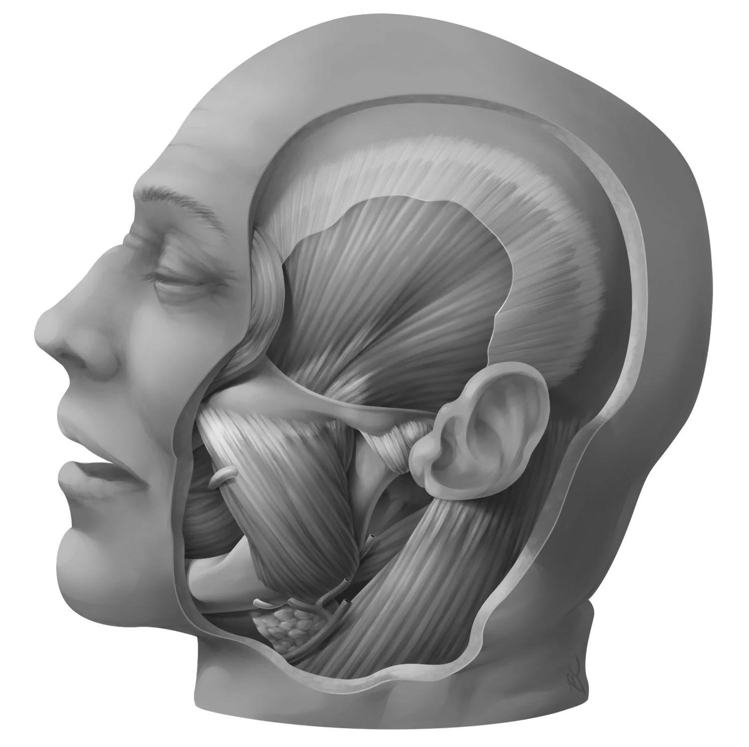
Grant’s Museum houses the dissections used to illustrate Grant’s Atlas, one of the most well-regarded anatomical atlases in the world. This piece was based on one of the specimens in the museum and demonstrates the ability to fuse real specimens, which can be messy and ambiguous, with information from other sources to create a clear and understandable representation.
Process Work
Sketching the Museum Specimen
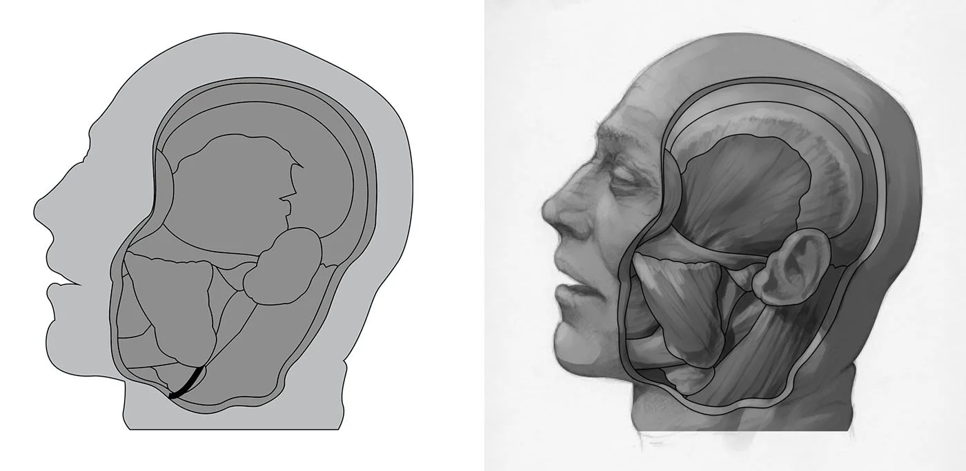
Since photos are not allowed inside Grant’s Museum, I sketched the museum specimen in as much detail as possible. On tracing paper, I sketched the major shapes and copied the labels provided by the museum’s diagram of the specimen.
Shapes and Preliminary Shading
I conducted research to ensure my drawing from the specimen was clear and anatomically accurate. Then, using Adobe Illustrator, I blocked out the major shapes. Afterwards, I went back to the museum and positioned a lamp to illuminate the specimen from the upper left and roughly shaded the illustration.
Rendering
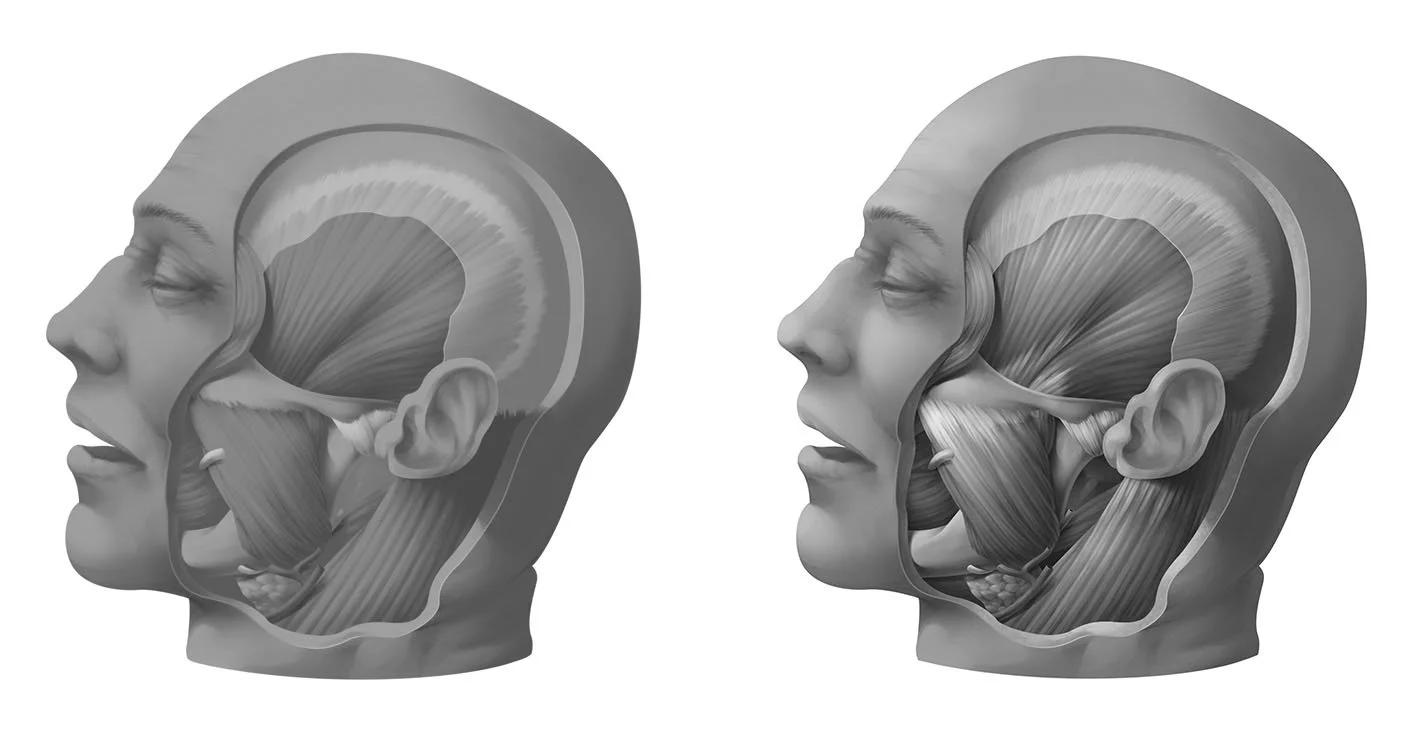
Using the preliminary shading from real life observation as a guide, I began to properly shade the illustration. Highlights, cast shadow, and texture were added afterwards. I added more detail and contrast in the area under the cut, since the exposed anatomy was the main focus. Labels were added in Adobe Illustrator after the illustration was complete.
References
Academic Sources
Agur, A. M. R., and Dalley, A. F. 2017. Grant’s Atlas of Anatomy, 14th ed. Philadelphia: Wolters Kluwer. Fig. 7.49: Temporalis and Masseter.
Gray, H. 2008. Gray’s Anatomy: The Anatomical Basis of Clinical Practice, 40th ed., ed. Standring, S. New York: Elsevier Churchill Livingstone. Fig 29.11: The superficial muscles of the head and neck and Fig 29.18: The principal immediate deep relations of the parotid gland.
Moore, K. L., and Dalley, A. F. 1999. Clinically Oriented Anatomy, 4th ed. Philadelphia: Lippincott Williams & Wilkins. Fig 7.6: Muscles of the scalp, auricle (external ear, pinna), face, and neck.
Visual References
Grant’s Museum. “H57A: Great muscles on the side of the skull”. University of Toronto. Accessed November 19, 2019.


